


Tooth in Eye Surgery: a Unique Approach
Imagine regaining sight with a lens crafted from your own tooth. This innovative procedure, known as osteo-odonto-keratoprosthesis (OOKP) or "tooth in eye" surgery, offers hope to individuals with severe corneal and ocular surface damage when traditional corneal transplants have failed or are not possible. In these cases, the cornea, the clear front part of the eye, becomes too cloudy or damaged to allow vision. OOKP creates a new window for vision using a remarkable technique.
The procedure, pioneered in the 1960s by Italian ophthalmic surgeon Professor Benedetto Strampelli, involves using the patient's own tooth to create a biocompatible support structure for an artificial cornea. Because the tissue is from the patient's own body, the risk of implant rejection is significantly reduced.
The Tooth in Eye Surgery Process
The "tooth in eye" surgery, or OOKP, is a complex, multi-stage procedure typically performed in two main stages. Here's a breakdown of the process:
Stage 1: Preparation and Grafting (Approximately 3 months)
- Tooth Removal and Preparation: Surgeons extract a canine or premolar tooth from the patient. This tooth is then meticulously shaped into a rectangular lamina.
- Lens Insertion: A hole is drilled into the prepared tooth lamina, and a plastic optical lens cylinder is inserted.
- Cheek Implantation: This tooth-lens complex is implanted into the patient's cheek. This crucial step allows the tooth to develop a new blood supply and supporting tissue, which is essential for the next stage of implantation in the eye.
- Eye Preparation: Simultaneously, the eye is prepared to receive the implant. This involves removing the damaged outer layer of the cornea and replacing it with a graft of soft tissue, often taken from the inner lining of the patient's cheek (buccal mucosa). This graft needs time to heal and integrate with the eye surface, typically a few months.
Stage 2: Eye Implantation
- Implant Extraction: After approximately three months, the tooth-lens complex, now with a layer of supporting tissue, is removed from the cheek.
- Eye Surgery and Implantation: The cheek mucosal lining over the eye is opened. The inner contents of the eye are removed to create space for the implant.
- Tooth-Lens Insertion: The prepared tooth-lens complex is carefully removed from the cheek and implanted into the eye. The mucosal cheek lining graft is then repositioned over the implant to secure it.
Following the surgery, light can now enter the eye through the implanted optical cylinder, potentially restoring vision for the patient. The entire process, from tooth removal to vision restoration, can take several months due to the tissue integration and healing phases.
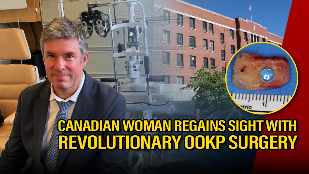
A tooth for an eye: Canadian Woman has Hope with this Innovative Eye Surgery
Earlier this week, at Mount Saint Joseph Hospital in Vancouver, Canada, Gail Lane underwent this rare operation, aka osteo-odonto-keratoprosthesis (OOKP). She was living in darkness for a decade, unable to see even your own reflection. Gail is now on a journey that could restore her sight, thanks to a groundbreaking procedure known as "tooth in eye" surgery. The whole operation and procedure was done by Dr. Greg Moloney, the ophthalmologist and surgeon at the hospital. "I haven’t seen myself for 10 years," Gail shared with a mix of nervousness and hope, "If I’m fortunate enough to get some sight back, there will be wonderful things to see." This innovative procedure, performed for decades worldwide but a first in Canada for this unique case, wherein used a patient's own tooth to construct a framework for a new artificial cornea.
Doctors carefully extracted one of Gail's teeth. This tooth wasn't destined for the tooth fairy, but for a much more extraordinary purpose. The tooth was meticulously shaved and shaped into a rectangle. A tiny hole was then drilled into it to house a custom-made plastic optical lens. This prepared tooth, now carrying a lens, was temporarily placed in Gail's cheek.
Dr. Moloney clarified that as the teeth lack the connective tissue needed for immediate attachment to the eyeball with sutures. The three-month stay in the cheek was crucial; as it allowed the tooth to develop a layer of supportive tissue, essentially becoming biologically integrated and ready for its new role in the eye. In preparation for the implant, Gail's eye also underwent a procedure. The top layer of the eye's surface was removed and replaced with a soft tissue graft from her cheek. This graft needed to heal fully before the "tooth lens" could be implanted.
This "tooth in eye" surgery offers a beacon of hope for individuals with corneal blindness. While still a rare and complex procedure, OOKP showcases the incredible advancements in modern ophthalmology and the potential to restore sight using the body's own remarkable resources. For Gail Lane, and others like her, this surgery is more than just a medical procedure; it's a chance to see the world anew.
DISCLAIMER: The information presented in this brief draws upon publicly available sources, including news reports, and industry publications, and expert commentary. The analysis and conclusions presented reflect the author's own understanding and perspective.